animal cell microscope experiment
A phagocytic cell can even engulf other structures. Investigating animal cells.

Microscope Lab Templates Plant And Animal Cells Science Projects For Kids Science Cells
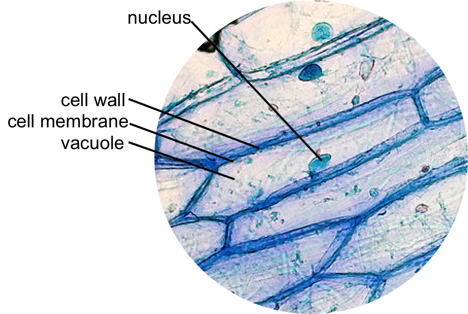
For this experiment the thin membrane will be used to observe the onion.

. Cultured cells and tissues will only behave normally in a physiological environment and control of factors such as temperature and tissue culture medium composition is thus critically important to obtaining meaningful data in. Biomedical Engineering Shared Laboratories. Many important advances in understanding cells have directly followed the development of new methods that have opened novel avenues of investigation.
An appreciation of the experimental tools available to the cell biologist is thus. You can make it as you. The cell from Latin word cellula meaning small room is the basic structural and functional unit of lifeEvery cell consists of a cytoplasm enclosed within a membrane which contains many biomolecules such as proteins and nucleic acids.
This animal cell 3d model helps us to learn about different parts of animal cell. An onion is made up of layers that are separated by a thin membrane. SlideShare uses cookies to improve functionality and performance and to provide you with relevant advertising.
In order to examine cells in the tip of an onion root a thin slice of the root is placed onto a microscope slide and. How to Obtain a Thin Layer of Onion Cells. We provide animal parentage verification identification coat color testing horse ancestry testing and genetic diagnostics of inherited disorders and traits.
This rap was created for a 6th-grade science classroom to teach about the different parts of a cell. One Day In 2022 Your Cell Phone May Kill You Or Turn You Into Zombie. Osmosis factors heavily in each of these processes and is an important force for keeping every single cell in your body healthy.
Due to the lack of a cell wall animal cells can transform into a variety of shapes. For instance whereas the nerve cells play a crucial role in the transmission of signals to different parts of the body blood cells play an important role carrying oxygen to different parts of the body. Most plant and animal cells are only visible under a light microscope with dimensions between 1 and 100 micrometres.
Osmosis is hard to see without a microscope. Today we are making animal cell model project for school. If you can isolate a single cell it.
Stained human cheek cells. We need powerful microscope for looking it. Animal cells are distinct from those of other eukaryotes most notably plants as they lack cell walls and chloroplasts and have smaller vacuoles.
All animals are eukaryotic. Here different types of cells play a specific function given that they have varied structures. Key parameters to consider before you start your live-cell imaging experiment.
Using this very simple staining procedure we can easily identify some of the basic structures of an animal cell. Animal cell culture technique is the process of developing cells invitro in a lab providing them invivo conditions. It is always best to image your live-cell samples using as little light as possible.
Basically there are two types of cells animal cell and plant cell. This animal cell working model is best for 7th grade science projects. Amoeba eats two paramecia.
Watch Woman Being Frequency Attacked By Her Cell Phone. Then Watch Kate Bush 80s Music Video Experiment IV About Frequency Sound Weapon Then Note Similarities To Cell Movie. Nuclei appear as small dark elliptical structures within the cell.
With its catchy rhythm and rhymes students of all learn. As in all experimental sciences research in cell biology depends on the laboratory methods that can be used to study cell structure and function. These regions of growth are good for studying the cell cycle because at any given time you can find cells that are undergoing mitosis.
Unlike animal cells such as cheek cells the cell wall of an onion and other plants are made up of cellulose which protects the cell and maintains its shape. Epidermal onion cells under a microscope. When planning a live-cell imaging experiment it is critical to devise an experimental plan that includes the important considerations highlighted below.
But if we create our very own model of a cell using a shell-less chicken egg we can see what happens when we manipulate the osmotic balance in the cell. The differences in structure and functions between the cells mean that they are specialized cells.

Onion And Cheek Cell Lab Experiment Organelles Science Cells Biology Classroom Life Science Middle School

Cells Microscope Activity Unit Microscope Activity Science Cells Science Lessons

Onion Cells Under A Microscope Requirements Preparation Observation Plant And Animal Cells Animal Cell Plant Cell

Cells Observation Lab Activity Plant Cell Lab Activities Cells Worksheet

Structure Of Animal Cell And Plant Cell Under Microscope Diagrams Cell Diagram Plant Cell Diagram Animal Cell

Cell Lesson Cheek And Onion Cell Cell Theory Cells Lesson Cell Theory Biology Units

Cells Microscope Things Under A Microscope Cell Plant And Animal Cells

Microscopic Animal Cells Images Kuhn Photo Microscopic Cells Microscopic Photography Things Under A Microscope

Epidermal Onion Cells Under A Microscope Plant Cells Appear Polygonal From The Cell Diagram Plant Cell Diagram Plant Cell